…Looking and not touching is called abandoning…
Thursday 1 February 2024

Breast pathology is becoming one of the most frequent topics of interest, either because of the symptoms or changes that we may experience or because of the situations we may be faced with.
So, we must remember that our mammary glands (“Breasts” – “Bubbies”) being present to give us a touch of beauty with their different sizes, or styles to carry, or feed and give growth and life support to our children, must be considered, cared for and watched over as an organ as important as our heart or kidneys among others. In this case I will also refer to the male gender, although they do not have food or beauty functions, they also present alterations and can generate pathologies. Therefore, the alterations presented in the mammary glands will not identify gender, age, race or beliefs (political or religious) and their spectrum of presentation will range from benign lesions such as fibroadenomas to malignant lesions known as breast cancer.
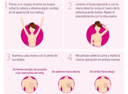
One of the pillars in the early detection of abnormalities in the mammary glands is self-examination, which is indicated to be performed every month. In women of childbearing age it is ideal to perform it 1 week after the menstrual period, in postmenopausal women at any time of the month. This is how it should be done:
And what are the warning signs in this self-examination that should lead us to consult a specialist?
- Masses or lumps in the breasts, armpits or above the collarbones.
- Hardening or swelling
- Redness or scaling of the skin or nipple
- Nipple engorgement or changes in position
- Nipple discharge during periods other than lactation
- Any change in the size or shape of the breasts
- Persistent pain anywhere in the breasts or armpits
- Swelling under the armpit or around the collarbone

Imagen tomada de www.cancerlatam.com
So if I have any of these warning signs I may have cancer, what is it and why does it happen?
If you have the presence of one or more of the warning signs, you should consult a specialist (mastologist or gynecologist) as a matter of priority to initiate a detailed evaluation and follow-up.
For the development of breast cancer we have risk factors that are related to the appearance of this pathology. Chek this link for more information. Within these we have modifiable and non-modifiable factors, as follows:
1. Modifiable risk factors:
- Sedentary lifestyle
- Overweight/obesity
- Hormone replacement therapy
- Late first pregnancy or no pregnancies
- Alcohol consumption
2. Non-modifiable risk factors:
- Age > 50 years
- Genetic mutations
- Early menarche, late menopause
- Dense mammary glandular tissue
- Personal history of breast cancer or previous breast pathologies such as “atypical hyperplasia” or “lobular carcinoma in situ”.
- Family history of breast cancer and/or ovarian cancer
- Previous treatments with radiotherapy
Breast cancer is considered as the abnormal and uncontrolled growth of mammary glandular cells, which originate a tumor and its growth has the capacity to invade adjacent regions or other organs, originating a process called “metastasis” (1).
Are there other surveillance tests besides the Self-Examination?
Yes, they are called screening tests; a clinical examination by a trained health professional is recommended. In women over 20 years of age, at least once every 3 years; and from 40 years of age onwards, once a year, or to any woman who consults for breast symptoms regardless of age.
In addition to the above two, control and surveillance with imaging (2) is indicated:
- Mammography: Indicated to be performed every two years in asymptomatic women between 50 and 69 years of age, and if life expectancy is greater than 10 years, continue to perform controls at this interval. But we must also know that there is another indication for requesting this study and it is when alterations are found in the clinical examination in women over 35 years of age.
- Breast ultrasound: Useful in diagnosis and follow-up of lesions in women under 35 years of age or with dense breasts, and allows differentiation between solid and cystic palpable and non-palpable lesions.
The respective report of each one of them is given in the BIRADS classification (3) and will allow to establish diagnostic suspicions and to direct a management.
- Breast MRI: Screening in high risk women (BRCA 1 and 2 mutations), patients with breast implants or to determine the extent of the disease in patients with already diagnosed breast cancer.
What is the current incidence of breast cancer?
It is the type of cancer with the highest incidence in the country and according to the WHO (4) in 2020 there were 2,261,419 new cases in the world and in Colombia it occupies the first place in prevalence with 13.7% of cancer in the country (5).

How is the diagnosis made and how is it managed?
Everything starts with diagnostic suspicion either by personal history, alterations in self-examination, clinical medical examination or incidental findings in screening images. Subsequently, the lesions are confirmed by means of various techniques that will allow the histological component to be evaluated (6). Among these techniques we find:
- Fine Needle Aspiration (FNA)
- Trucut needle biopsy
- Stereotaxy biopsy
Then, according to the specific histology report, the nature of the lesion, benign or malignant, can be defined and management can begin.
Management will not only depend on the unique characteristics of the lesion, since if malignant lesions are found, additional extension studies should be performed to evaluate (confirm or rule out) the presence of cancer outside the breast.
Management (7) will have several starting and follow-up points, as follows:
- Observation
- Surgical management (quadrantectomy, mastectomy)
- Pharmacological management
- Radiotherapy
- Palliative management
Therefore, after this quick review of breast pathology, basically breast cancer, we must reflect that “look and don’t touch is called neglect” because not properly monitoring and neglecting our “Breasts” can lead us to drastically change the course of our lives and the loved ones around us.
References
- CDC en español
- Diaz, S.E et al (2015) Manual for early detection of breast cancer. Pages 22-27. Retrieved from
- Camacho, C (2018) Update of the BI-RADS nomenclature by mastography and ultrasound.
Annals of Radiology Mexico.
Retrieved from - WHO en español
- Liga Contra el Cáncer Colombia
- Diaz, S.E et al (2015) Manual for early detection of breast cancer. Pages 24-27. Retrieved from
- Diaz, S.E et al (2015) Manual for early detection of breast cancer. Pages 40-50. Retrieved from


